Visual cues determine hippocampal directional selectivity
Lavanya Acharya, Zahra M. Aghajan, Cliff Vuong, Jason J. Moore, Mayank R. Mehta

TL;DR
This study shows that visual cues alone can drive directional selectivity in rodent hippocampal neurons, challenging previous assumptions about the role of vestibular input.
Contribution
The study demonstrates that visual cues are sufficient and causal in driving hippocampal directional selectivity, even without strong vestibular input.
Findings
Hippocampal neurons show significant head-direction modulation during random foraging in real-world environments.
Directional modulation in hippocampal neurons is also observed in virtual reality with minimal vestibular input.
Changes in visual cues directly affect hippocampal directional modulation, with ensemble activity biased toward specific visual cues.
Abstract
Both spatial and directional information are necessary for navigation. Rodent hippocampal neurons show spatial selectivity in all environments1, but directional tuning only on linear paths2–8.The sensory mechanisms underlying directionality are unknown, though vestibular and visual cues are thought to be crucial. However, hippocampal neurons are thought to show no angular modulation during two-dimensional random foraging despite the presence of vestibular and visual cues6,7. Additionally, specific aspects of visual cues have not been directly linked to hippocampal responses in rodents. To resolve these issues we manipulated vestibular and visual cues in a series of experiments. We first measured hippocampal activity during random foraging in real world (RW) where we found that neurons’ firing exhibited significant modulation by head-direction. In fact, the fraction of modulated neurons…
Click any figure to enlarge with its caption.
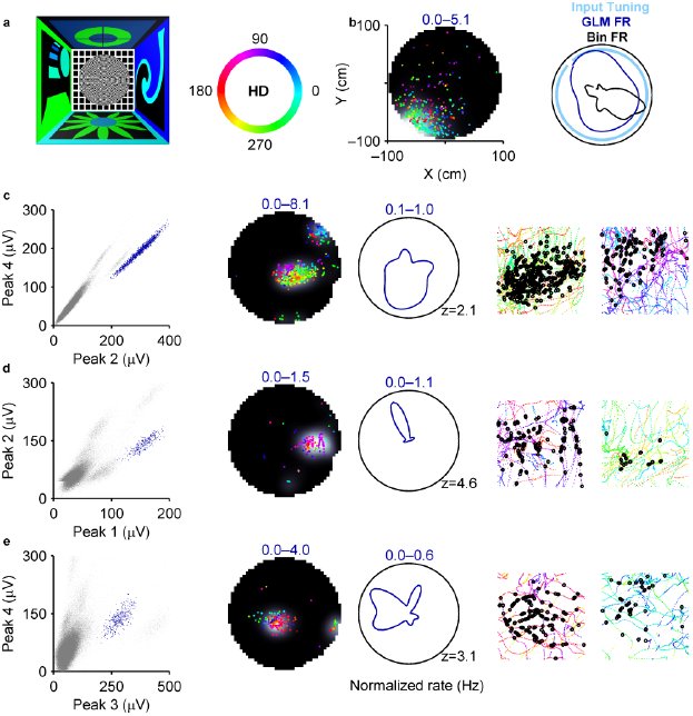 Figure 1
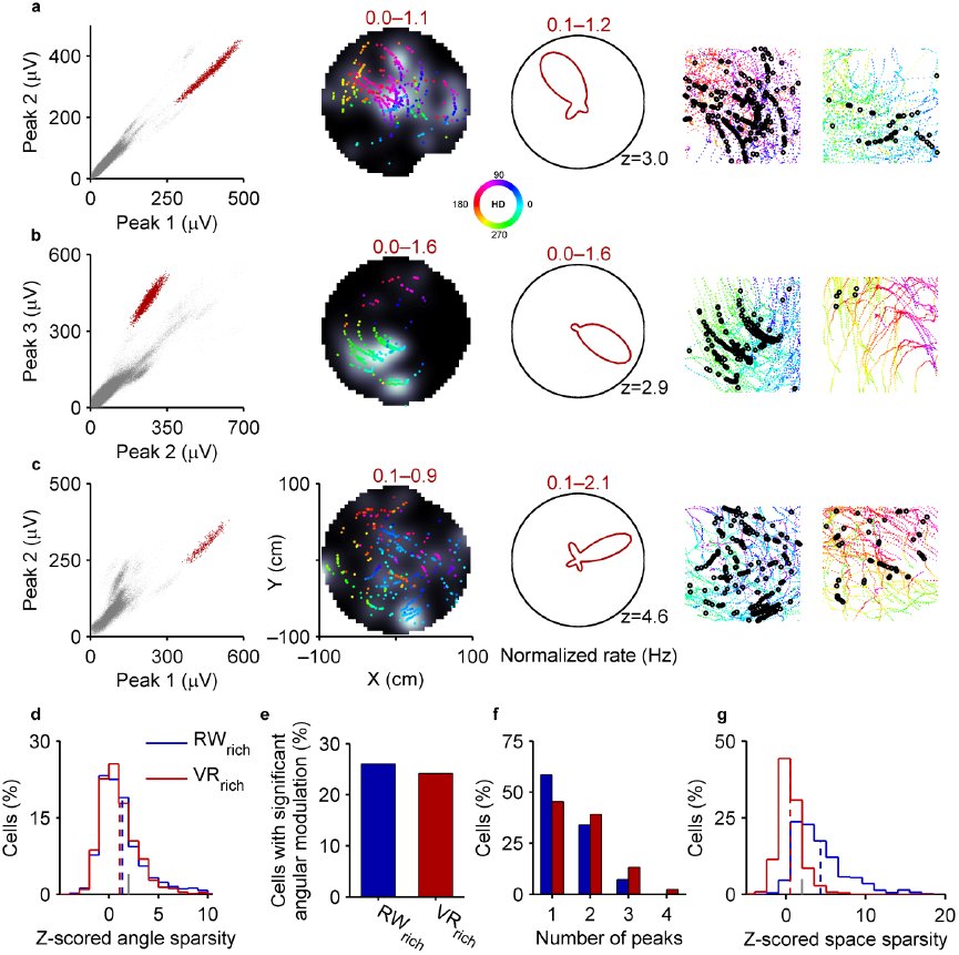 Figure 2
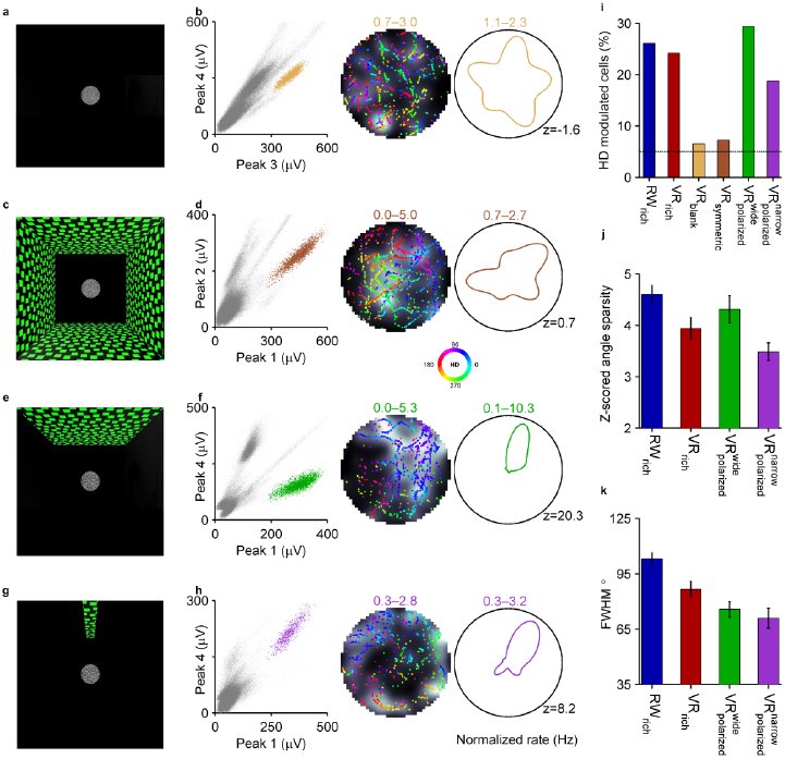 Figure 3
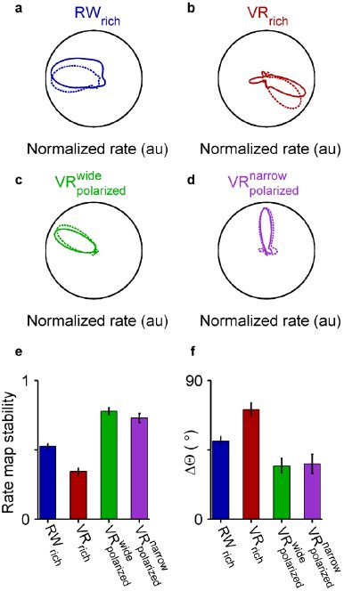 Figure 4
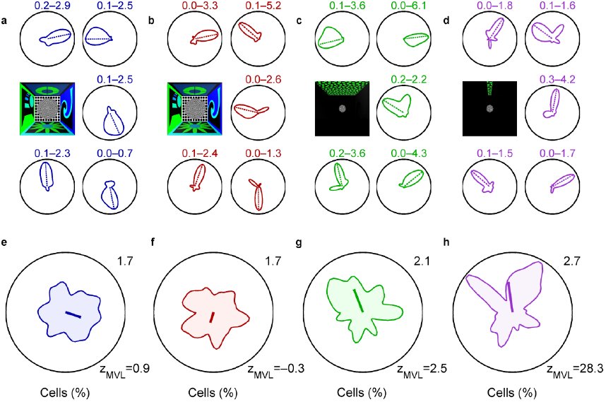 Figure 5


















Peer Reviews
No public reviews on file for this paper yet. If you reviewed it on a platform where reviews are public (OpenReview, ICLR, NeurIPS, ICML), you can paste yours below so the community can read it here.
Videos
No videos yet. Explain this paper in a talk, walkthrough, or lecture? Add one.
Taxonomy
TopicsMemory and Neural Mechanisms · Neuroscience and Neuropharmacology Research · Categorization, perception, and language
Introduction
Hippocampal spatial selectivity has been well established and the underlying mechanisms extensively studied^1,12^. However, both the existence and mechanisms of hippocampal directional selectivity are debated. During random foraging in two-dimensional arenas, barring a few conflicting reports^3,6,13^, the consensus is that rodent hippocampal neurons do not show significant angular selectivity^6,7^. In contrast, hippocampal neurons exhibit strong directional selectivity on linear paths^3–5,8^. The reason for this disparity and the sensory mechanisms of directionality are unclear, although visual and vestibular cues have been proposed as likely candidates^3,5,14^. In addition, internal mechanisms also contribute to hippocampal activity^11,15–17^.
Visual cues strongly influence the spatial firing properties of hippocampal neurons ^2,18^. Further, comparable levels of directionality exist on linear tracks in RW and in VR^5^ where the range of vestibular inputs is minimal, suggesting that visual cues also influence directionality in one dimension. In addition, selectivity to the visual cue towards which the animal’s head is facing, referred to as spatial-view, has been reported in humans^19^, primates^20,21^ and bats^22^. However, response to specific features of visual cues has not been observed in rodents, leading to the notion that in these animals visual cues merely provide a context for hippocampal activity.
In parallel, vestibular inputs are crucial to the head-direction system, which is thought to provide directional information to the hippocampus. Consistently, vestibular lesions disrupt hippocampal spatial selectivity^23^, although lesions in the head-direction system do not^24^. However, if instantaneous vestibular cues were contributing to hippocampal directionality, there should be greater directionality in two-dimensional RW tasks, where the range is higher compared to one-dimensional RW tasks, but the opposite is true. Some studies have attributed directionality in two dimensions to vestibular-derived self-motion information^3,14,25^, but no study, to our knowledge, has directly measured hippocampal head-directional modulation when vestibular-based signals are impaired.
Thus, the mechanisms governing hippocampal directional activity in rodents are unclear. We hypothesize that visual cues directly influence the activity of hippocampal neurons to generate angular tuning whereas vestibular cues are not required for directionality.
Results
To test these hypotheses we did a series of experiments and analyses. We first quantified hippocampal spatial and head-directional modulation from 1066 active (defined as cells with minimum mean firing rate of 0.2Hz and with at least a 100 spikes) dorsal CA1 pyramidal neurons (which were part of a previous study of hippocampal spatial selectivity^11^). Rats randomly foraged for rewards on a two-dimensional platform in a RW environment which had rich distal visual cues and will henceforth be referred to as RW_rich_ (Fig. 1a).
Presence of head-directional modulation in hippocampal pyramidal neurons in RWrich. a, Left) A top-view schematic depicting a 300cm×300cm room with four different visual cues on the walls and an elevated 100cm radius platform at the center. Right) A color wheel representing the mapping between head-directions and colors. b, Left) Spatial firing rate of a surrogate neuron (grey scale range indicated by numbers; lighter shades correspond to higher values, here and throughout all figures) overlaid with the position of the rat when spikes occurred (colored dots). Each color represents a distinct head-direction as shown in (a). The surrogate neuron’s activity was constructed to have significant spatial selectivity but no angular selectivity. Right) Angular rate map of the same surrogate neuron estimated using the binning (black) and GLM (blue) methods, along with the uniform input tuning (light blue). The GLM method provided an accurate estimate of the input, but the binning method overestimated the angular tuning due to behavioral bias (Extended Data Fig. 1). c, Left) All unclustered (grey dots) and clustered spike amplitudes from an isolated neuron (blue dots) on two different projections of a tetrode in RWrich. Center) Spatial and angular rate maps of a cell (same convention as in (b)). Numbers in color indicate range, here and throughout unless noted otherwise. The number at the bottom right corner of the polar plot is the z-scored sparsity of the angular rate map). Right) Rat’s color-coded trajectory and his position at the time of spikes (black circles) for movement in the direction of maximal (left) and minimal (right) firing respectively. d,e, Same as (c) for two other cells in RWrich. All rate maps were computed using the GLM method here and throughout unless otherwise noted.
A common technique for quantifying head-directional modulation is to divide the number of spikes in each direction bin by the total time spent in that bin (Fig. 1b)^9^. However, when neurons have spatially tuned responses, as is the case for hippocampal neurons in RW, this method provides incorrect estimates of angular tuning^6^. For example, for a neuron with a place field at the edge of the maze, this method would yield artificially large head-directional tuning due to non-uniform sampling of head angles within the place field (Fig. 1b, Extended Data Fig. 1)^6^. Various methods have been developed to overcome this confound^3,25,26^. Here, we adopted the well-established generalized linear model (GLM) approach (see Methods)^15,27–29^ which has several advantages. First, it provides an unbiased estimate of the simultaneous and independent contribution of spatial and head-directional modulation. Second, unlike other methods, head-directional modulation obtained with the GLM method is uninfluenced by behavioral biases within the place field as verified using surrogate data with predetermined levels of spatial and angular modulation (see Methods, Extended Data Fig. 1i). Finally, this method provides an estimate of the fine structure of the respective tuning curves.
This method revealed a surprising finding: many neurons exhibited clear modulation by the rat’s head-direction in RW_rich_(Fig. 1c,d,e, Extended Data Fig. 2a, see Methods). Some neurons fired maximally for only one head-direction and minimally elsewhere (Fig. 1c,d), while others showed a multimodal response (Fig. 1e). The statistical significance of head-directional modulation was quantified by the sparsity of angular rate maps z-scored with respect to control data (see Methods). Angular sparsity thus defined was significantly (p<0.05) greater than chance (>2z) for 26% of neurons in RW_rich_. In fact, the fraction of hippocampal neurons with significant angular tuning was comparable to that in many parts of the head-direction system, although the width of the angular tuning curves (full width at half maximum 103.10±3.50°) was wider^9,30^.
Similar levels of significant head-directional modulation in RWrich and VRrich. a–c Three well-isolated neurons showing significant head-directional modulation in VRrich (same conventions as in Fig. 1). d, Z-scored angular sparsity in VRrich (1.09±0.08, n=719, red) was similar (p=0.2) to that in RWrich (1.35±0.08, n=1066, blue). Mean values are indicated with dashed vertical lines. Grey tick mark indicates significance threshold (z=2). e, 26% (24%) of cells in RWrich (VRrich) showed significant head-directional modulation (see Methods). f, Head-directionally modulated neurons in VRrich were significantly more multimodal (1.72±0.06 peaks, p=1.5×10⁻³) than RWrich cells (1.49±0.04 peaks). g, In contrast to similar z-scored angular sparsity, z-scored spatial sparsity was significantly reduced in VRrich (0.51±0.06, p=1.2×10−174) compared to RWrich (4.32±0.11). Throughout the figure legends values are reported as mean±s.e.m and the statistical significance for comparisons was computed using Wilcoxon rank-sum test unless otherwise stated.
This raises an important question: which sensory inputs could generate the head-directional modulation in our data? Two likely candidates are the visual and vestibular modalities. To dissociate the two, we measured the activity of 719 active dorsal hippocampal CA1 pyramidal neurons^11^ during the same random foraging task in a two-dimensional VR environment (VR_rich_). Here, the distal visual cues were identical to those in RW_rich_, but the range of vestibular cues was minimized due to body fixation. Despite impaired spatial selectivity^11^, many neurons showed clear modulation by the direction of the rat’s head with respect to the distal visual cues, which will be henceforth referred to as “head-direction"(Fig.2a,b,c, Extended Data Fig. 2b).
Curiously, across the ensemble of neurons there was no substantial difference in head-directional modulation between the two worlds as quantified by z-scored angular sparsity (Fig. 2d). Further, the fraction of neurons showing significant directional modulation in VR_rich_ (24%) was similar to that in RW_rich_ (Fig. 2e). Neurons in VR_rich_ also had multimodal responses like in RW_rich_, unlike neurons in the head-direction network which have unimodal responses^31^. The multimodality was greater in VR_rich_ than RW_rich_ (Fig. 2f) which could account for slightly lower z-scored mean vector length in the former (Extended Data Fig. 3). This observation motivates the use of z-scored sparsity as a measure for angular selectivity. Additionally, the width of the angular tuning curves in VR_rich_ (86.61±4.19°) was significantly (16%, p=4.3×10^−4^) sharper than in RW_rich_.
Direct influence of visual cues on the degree of directional modulation of neurons a, Top-down schematic of VR task with a 100cm radius circular platform with no distal visual cues (VRblank). b, Left) Spikes from an isolated neuron (mustard dots) in VRblank (same convention as in Fig. 1). Center, right) Spatial and angular firing rate of this neuron (same conventions as in Fig. 1). c, Top-down schematic of VR task with symmetric cues located 450cm away from the center of the circular platform (VRsymmetric). d, Same as in (b) but in VRsymmetric. Note that the neurons shown in both (b, d) do not exhibit any head-directional modulation (z<2). e, Top-down schematic of VR task with a single wide polarizing visual cue 450cm away from the center (90° visual angle) of the platform . f, Same as (b, d) but in Top-down schematic of VR task with a narrow polarizing cue 450cm away from the center (11° visual angle) of the circular table . h, Same as (b, d, f) but in . Note that the neurons shown in both (f, h) exhibit strong head-directional modulation (z>2). i, The percentages of cells with significant head-directional modulation was 26% in RWrich (278 out of 1066 cells), 24% in VRrich (174 of 719 cells), 6% in VRblank (14 of 230 cells), 7% in VRsymmetric (30 of 426 cells), 30% in (89 of 300 cells) and 19% in (65 of 341 cells). The black horizontal line indicates the chance level of 5%. The distribution of z-scored angular sparsity was significantly different from zero in RWrich (p=3.0×10−59 Wilcoxon signed-rank test here and throughout (i)), VRrich (p=3.0×10−37), and but not in VRblank(p=0.7) or VRsymmetric (p=0.2). j, Z-scored sparsity of the angular rate maps for cells with significant head-directional modulation in different tasks was 4.61±0.17 in RWrich, 3.94±0.21 in VRrich, 4.31±0.26 in and 3.49±0.17 in (error bars indicate s.e.m). Out of six possible pair-wise comparisons between the distributions, the only significant differences were that z-scored angular sparsity in RWrich was slightly greater than VRrich (p=4.1×10⁻³) and . k, Full width at half max (FWHM) of the angular rate maps for head-directionally modulated neurons in different conditions was as follows: RWrich (103.10±3.50°), VRrich (86.61±4.19°), and . The tuning curves in RWrich were significantly wider than all other VR conditions (p=4.3×10⁻⁴ versus VRrich, p=4.9×10⁻⁵ versus and p=6.8×10⁻⁶ versus ). Within VR conditions, the only significant difference was polarized polarized polarized observed between VRrich and with the latter having significantly narrower tuning curves (p=0.03). Values are reported as mean±s.e.m and the p-values are obtained by Wilcoxon rank-sum test unless noted otherwise.
We then quantified the spatial modulation of neural responses in both RW_rich_ and VR_rich_ using the rate maps obtained from the GLM method. We found significant spatial selectivity in RW_rich_ but not in VR_rich_ (Fig. 2g), consistent with previous results obtained using the binning method^11^. Although head-directional modulation was comparable between the two worlds, spatial modulation was not, which suggests a decoupling of the mechanisms of spatial and directional tuning. Consistently, the presence or absence of head-directional modulation had no effect on the percentage of spatially modulated neurons in both RW_rich_ and VR_rich_(Extended Data Fig. 4a).
Stability of angular rate maps for head-directionally modulated neurons. a–d, Four directionally tuned cells with stable angular firing in the first half (solid colored lines) and second half (dashed colored lines) of the recording session. The peak rates are normalized for ease of comparison. e, Stability of the head-directional modulation (computed as the pairwise correlation between the angular rate maps in the first and second halves) in RWrich (0.53±0.02, n=278) was significantly greater than VRrich (0.34±0.02, n=174, p=2.2×10⁻⁹, Wilcoxon rank-sum test here and throughout figure legend) but significantly smaller than (0.78±0.02, n=89, p=1.3×10⁻¹⁶) and (0.73±0.03, n=65, p=3.2×10⁻⁹). Angular rate map stability was not significantly different between and (p=0.2). f, As an alternate measure of stability, we computed the absolute value of the circular distance between the preferred directions (defined as the direction of peak firing) in the two session halves. This method also resulted in a similar trend with (34.59±4.75°) and (35.89±6.19°) showing identical levels of drift of the preferred directions (p=0.5), both smaller than the amount of angular drift in RWrich (50.78±2.94°, p=4.3×10⁻⁵ and p=1.1×10⁻⁴ respectively) and VRrich (71.16±4.23°, p=1.4×10⁻⁸ and p=4.7×10⁻⁷ respectively).
Interestingly, directionally tuned neurons had greater mean firing rates than the untuned neurons in VR_rich_ (Extended Data Fig. 4b). This difference was not present in RW_rich_, which might be due to the presence of multisensory spatially informative cues^11^.
These results show that rodent hippocampal neurons in RW indeed show significant head-directional modulation during two-dimensional random foraging, contrary to previous reports. In addition, the observation that head-directional modulation remained intact in VR—where the range of vestibularcues is minimized—suggests that vestibular cues are not required for hippocampal head-directional modulation.
What other mechanism could generate angular modulation? Either it is internally generated^15–17^ or driven by specific visual cues^20,21^. To disambiguate these possibilities, we generated a virtual world where distal visual cues were entirely eliminated (VR_blank_)(Fig. 3a, see Methods). The circular platform in the virtual environment, which was the only visual cue present, provided optic flow information but had no spatial or angular information. The rats’ behavior in VR_blank_ was comparable to that in VR_rich_ with visually distinct walls (Extended Data Fig. 5a). Hippocampal neurons did not show clear head-directional modulation in this case (Fig. 3b,i, Extended Data Fig. 5b) and the distribution of z-scored angular sparsity was not significantly different from zero (p=0.7, Wilcoxon signed-rank test).
Bias of the neural ensemble by polarizing visual cues. a, Five example cells in RWrich with significant directional tuning. The numbers indicate firing rate range. The dashed line corresponds to the preferred direction, i.e. the direction of maximum firing. The schematic in the middle left indicates the experimental condition. b–d, Same as in (a) but in VRrich, and conditions respectively. e, Distribution of preferred direction of neurons in RWrich(341.71±4.66°, n=278, circular mean±circular s.e.m). The number on the top right indicates the maximum value of the distribution. The thick blue line originating at the center of the polar plot represents both the direction (341.71°) and the magnitude (0.08) of the mean vector length of the preferred directions of the population (scaled by a factor of 5 for clarity). The mean vector length was not significantly different from chance (z=0.95). f, Same as in (e) but for VRrich. The distribution of preferred directions of neurons in VRrich (253.27±5.97°, n=174, circular mean±circular s.e.m) was not significantly different from that in RWrich (p=1, circular Kuiper test). The mean vector length of the population (0.05) was not significantly different from chance (z=-0.34). g, The ensemble of head-directionally modulated neurons in , circular mean±circular s.e.m) were preferentially firing towards the visual cue (p=0.04, circular V test). Polarized Note the direction (111.27°) and the longer magnitude (0.12, z=2.50 which is significantly greater than chance) of the thick green line compared to (e, f). h, On the population level, neurons in , circular mean±circular s.e.m) were most biased towards the narrow cue (p=0.04, circular V test) as indicated by the magnitude (0.15, z=28.32) of the thick purple compared to all other conditions.
The absence of head-directional modulation in VR_blank_ may result from a lack of anchoring visual cues^32^ or optic flow created by the distal visual cues which could potentially be integrated to generate directional tuning. To address this, we performed another experiment where all the virtual walls had the same visual texture with high contrast and spatial frequency (VR_symmetric_), thus providing strong optic flow information but no angular information (Fig. 3c, see Methods). The virtual platform was placed in a larger room where each wall was 450cm away from the platform center, which ensured that the distance from the walls provided minimal spatial and angular information. Here too, neurons did not exhibit head-directional modulation (p=0.2, Wilcoxon signed-rank test), similar to VR_blank_ (Fig. 3d,i, Extended Data Fig. 5b). In fact, there was no significant difference between the degrees of angular modulation in VR_blank_ and VR_symmetric_ (p=0.6, Wilcoxon rank-sum test).
While internal mechanisms and optic flow may still modulate the degree of angular tuning, these experiments show that directional modulation is not generated by these mechanisms alone. This leaves open the possibility that head-directional modulation is generated by the angular information contained in the distal visual cues. To confirm this hypothesis we performed another experiment where the virtual world was strongly visually polarized. In this condition, there was just one high contrast wall, 450cm from the center of the platform, subtending a 90 degree angle [eqn](Fig. 3e, see Methods). This polarizing cue had no other spatial information and was identical to the walls used in the symmetric world. Here, 30% of hippocampal neurons showed robust head-directional modulation (Fig. 3f,i, Extended Data Fig. 6), which is a greater fraction than in all other conditions. However, the z-scored angular sparsity of neurons in [eqn] was comparable to that in RW_rich_ and VR_rich_ (Fig. 3j). Remarkably, the directional tuning curves of many neurons were much narrower (75.69±4.18°) than the sole, 90° wide polarizing cue.
Is there a lower bound on the width of the angular tuning curves? To address this we conducted another experiment where the sole polarizing cue was very narrow (11°), thus providing high angular information in the direction of the cue while leaving the majority of the maze blank [eqn](Fig. 3g, see Methods). Strong head-directional tuning was found in this condition as well (Fig. 3h, Extended Data Fig. 7). For neurons with significant head-directional tuning, the width of the tuning curves (70.94±5.34°) was not much narrower than in [eqn] (Fig. 3k), and much wider than the 11° polarizing cue, indicating a lower bound on the width of hippocampal angular tuning curves. This could be influenced by internally generated hippocampal motifs enforcing a lower bound on the duration of the firing and hence the width of the tuning curves^11^. Further, the fraction of neurons showing significant head-directional modulation (19%) was considerably lower than in [eqn] (Fig. 3i), perhaps because the narrow visual cue is visible to the rat for a smaller fraction of time than the wider polarizing cue, hence modulating a smaller percentage of neurons. Notably, despite similar z-scored angular sparsity for neurons with significant head-directional modulation in different conditions, z-scored mean vector length exhibited a different pattern which can be explained by the differences in the multimodality of the angular rate maps (Extended Data Fig. 8).
We then asked if the head-directional modulation of hippocampal neurons is stable and whether the stability depends on the experimental condition (Fig. 4). Tuning curves of neurons with significant head-directional modulation were significantly stable across the experimental session in all four conditions (RW_rich_ p=1.3×10^−43^, VR_rich_ p=2.0×10^−22^, [eqn] p=4.4×10^−16^ and [eqn] p=9.2×10^−12^, Wilcoxon signed-rank test)(Fig. 4a–d). The tuning curves were more stable (p=2.2×10^−9^) in RW_rich_ than in VR_rich_ (Fig. 4e,f). This could be due to the presence of other directionally informative multisensory cues in RW, such as distal odors and sounds, and their consistent pairing with visual cues resulting in higher stability. On the other hand, the tuning curves were more stable in the polarized VR experiments than in either of the rich conditions (Fig. 4e,f) indicating there may be competing influences of multiple cues within each modality in the rich conditions.
These results demonstrate that specific aspects of visual cues modulate the angular tuning of individual neurons; could they also influence the ensemble response? To address this we investigated the activity of the head-directionally modulated neurons on a population level under the four different conditions. For each neuron, the direction of maximum firing was computed from its angular rate map and was designated as its preferred direction (Fig. 5a–d, see Methods). We then computed the distribution of these preferred directions and the degree of angular bias of the population for each condition. There was no significant angular bias, as measured by z-scored mean vector length of the population (see Methods), in both RW_rich_ (z=0.95) and VR_rich_ (z=–0.34), and the two distributions were not significantly different from each other (p=1, circular Kuiper test) (Fig. 5e,f). The lack of population bias in the rich conditions is likely due to the presence of multiple visual cues on all walls, each contributing to tuning towards different directions.
Indeed, in [eqn] with only one visual cue, the population was significantly biased (z=2.50). This directional bias of the population was significantly (p=0.04, circular V test) oriented towards the prominent visual cue (Fig. 5g). The directional bias of the population was even stronger in [eqn], and was also significantly (p=0.04) oriented towards the narrow visual cue. There was an apparent reduction in the number of cells with preferred direction directly towards the narrow polarizing cue, and for some cells the preferred direction was opposite to the visual cue. This suggests that while visual cues drive the angular selectivity of hippocampal neurons, network mechanisms can modulate this activity.
Discussion
These results demonstrate that, during two-dimensional random foraging, rodent hippocampal CA1 neurons show significant modulation as a function of head-direction with respect to the surrounding distal visual cues in both real and virtual worlds. This directional modulation does not require robust vestibular cues, while angularly informative visual cues are sufficient for its generation. Additionally, the directional modulation is strongly influenced by specific aspects of the distal visual cues, both at the neuronal and ensemble level.
Our demonstration of significant head-directional modulation of hippocampal neurons’ activity during random foraging in two dimensions in RW is contrary to the commonly held belief that head-directional modulation is absent in this condition^6,7^. The few reports about directionally modulated activity in hippocampus have reached conflicting conclusions, with some reporting vestibular-based head-directional modulation^3,14,25^ and others reporting vision-based spatial-view modulation^20–22^. By utilizing a VR setup we were able to isolate the contribution of only distal visual cues to hippocampal directional modulation. In addition, our analysis method was able to estimate the independent contribution of position and head-direction to hippocampal activity with high resolution, uninfluenced by behavioral biases^6^.
If hippocampal directional tuning was generated primarily from vestibular-based signals, one would expect a dramatic reduction of head-directional modulation in VR where the range of vestibular cues is minimized. However, hippocampal neurons not only showed significant levels of head-directional modulation in VR, but was also exhibited comparable levels to those in RW, thus demonstrating that robust vestibular cues are not necessary to generate directional tuning and that distal visual cues are sufficient.
About a quarter of all active CA1 neurons showed significant head-directional modulation in the visually rich RW and VR conditions, which is comparable to the fraction of directionally tuned neurons in several parts of the head-direction system^9^. We hypothesize that the directionally modulated hippocampal neurons may be the subset of the population that is predominantly driven by distal sensory cues, including distal visual cues. The rest could be largely driven by proximal cues, such as textures and odors on the track, which are not directionally informative. This hypothesis is consistent with prior studies showing a reduction in directional activity in the hippocampus with the inclusion of proximal cues^4^, and the remapping of subsets of hippocampal cells in accordance with the rotation of either distal or proximal cues^2,18,33,34^.
Head-directional modulation was abolished in VR in conditions with either no distal visual cues or symmetric distal visual cues demonstrating that it is not generated by internal mechanisms or optic flow alone. The directional modulation reappeared in VR with visually polarizing cues indicating that the angular information provided by visual cues directly influences head-directional modulation of individual hippocampal neurons. This influence was also observed at an ensemble level where the angular bias of the population was directly governed by the visual cues, i.e. no ensemble bias in the visually rich environments but a significant bias in the visually polarized cue conditions. Further, the degree of angular bias of the ensemble increased with increasing degree of angular polarization of the visual cue. Finally, this ensemble bias was pointed towards the polarizing visual cue. These results demonstrate a causal role of distal visual cues in driving hippocampal head-directional modulation.
Although a smaller fraction of neurons exhibited significant angular modulation in [eqn] compared to the rich conditions, the degree of ensemble bias showed the opposite trend. We speculate that this is because, in the rich conditions, different neurons fire preferentially to different visual features on the walls, resulting in a greater fraction of angularly modulated cells and no ensemble bias. In contrast, in [eqn] the visual features modulating neural responses were concentrated within a small range of angles, resulting in a reduction of fraction of directionally tuned neurons but a greater bias of the ensemble. These results indicate that visual cues do not provide a mere context for hippocampal activation, but rather that specific aspects of visual cues play a direct role in determining hippocampal responses.
Visual cues influence directional modulation of neurons in the head-direction system as well^9,35^. However, those neurons lose their direction selectivity without robust vestibular cues^36^, which is not the case in our hippocampal data from VR. Hence, we hypothesize that while the hippocampus may receive directional signals from the head-direction system, it must also be receiving directional information from a pathway that does not require the vestibular signal. This pathway could be through the parietal and retrosplenial cortices, which project to the hippocampus via the entorhinal cortex^9,37^ where neurons show significant head-directional modulation^38,39^.
In addition to the independence from vestibular cues, there are also other major differences between the properties of angular tuning curves in hippocampus and head-direction system. First, in hippocampus the tuning curves are much broader and more multimodal than those in the head-direction system^9,30^. Second, in the presence of a single polarizing visual cue, CA1 ensemble activity manifests a large bias in the direction of the visual cue, unlike the uniform distribution of preferred orientations in the head-direction system^31^.
Notably, the large reduction in spatial selectivity in VR^11^ is in contrast to the intact head-directional modulation observed here. This suggests that the mechanisms of spatial and directional selectivity can be dissociated, in that visual cues are sufficient to generate the latter but not the former. Further, these results bridge the gap between rodent, human and non-human primate studies showing the presence of angular selectivity independent of spatial selectivity^19,21,40,41^.
Our findings also narrow the gap between the presence of directionality on linear tracks^8^, but its apparent absence during random foraging in two dimensions^6,7^. Visual cues could generate the directional modulation of neurons in both one and two dimensions, and consistent pairing of visual and locomotion cues^11^ could enhance the degree of directional modulation on linear paths^3^.
These results could potentially resolve the apparent paradox: If the hippocampus is required for navigation^42^, how can rats navigate in a virtual world^10^ without hippocampal spatial selectivity^11^? We hypothesize that angular selectivity of hippocampal neurons, combined with their selectivity to distance traveled^5,11^, may be sufficient to solve a navigation task.
Methods summary
Five adult male Long-Evans rats foraged for randomly scattered rewards in RW and various VR tasks. Four rats ran in visually similar RW and VR tasks with environments identical in size (300cm×300cm room with a 100cm radius elevated circular platform at the center). In addition, two rats ran on a 100cm radius platform in four other VR tasks with different distal visual features to determine their influence of hippocampal firing as follows: a) a room with no distal visual cues; b) a room with angularly symmetric cues with high spatial contrast positioned 450cm away from the center; c) a similar environment as in (b) but with only one high contrast cue subtending a visual angle of 90° at the center; d) a similar environment as in (c) but with the visual cue subtending only 11° angle at the center. Electrophysiological data were collected using bilateral hyperdrives with 22 tetrodes from dorsal CA1^5,11^. All procedures were in accordance with NIH approved protocols. Spatial and head-directional modulations were computed using a generalized linear model framework^15,27–29^. See online methods for details.
The reference list from the paper itself. Each links out to its DOI / PubMed record.
- 1O’Keefe, J. & Dostrovsky, J. The hippocampus as a spatial map. Preliminary evidence from unit activity in the freely-moving rat. Brain Res. 34, 171–5 (1971).
- 2Muller, R. U. & Kubie, J. L. The effects of changes in the environment on the spatial firing of hippocampal complex-spike cells. J Neurosci 7, 1951–68. (1987).
- 3Markus, E. J. et al. Interactions between location and task affect the spatial and directional firing of hippocampal neurons. J. Neurosci. 15, 7079–94 (1995).
- 4Battaglia, F. P., Sutherland, G. R. & Mc Naughton, B. L. Local sensory cues and place cell directionality: additional evidence of prospective coding in the hippocampus. J Neurosci 24, 4541–4550 (2004).
- 5Ravassard, P. et al. Multisensory control of hippocampal spatiotemporal selectivity. Science 340, 1342–6 (2013).
- 6Muller, R., Bostock, E., Taube, J. & Kubie, J. On the directional firing properties of hippocampal place cells. J. Neurosci. 14, 7235–7251 (1994).
- 7Andersen, P., Morris, R., Amaral, D., Bliss, T. & O’Keefe, J. The hippocampus book. (Oxford University Press, 2006).
- 8Mc Naughton, B. L., Barnes, C. A. & O’Keefe, J. The contributions of position, direction, and velocity to single unit activity in the hippocampus of freely-moving rats. Exp Brain Res 52, 41–49 (1983).