Physisorption of Nucleobases on Graphene
S. Gowtham, Ralph H. Scheicher, Rajeev Ahuja, Ravindra Pandey, Shashi, P. Karna

TL;DR
This study uses first-principles calculations to analyze how different nucleobases interact with graphene, revealing varying binding strengths primarily driven by molecular polarizability, advancing understanding of DNA-carbon nanostructure interactions.
Contribution
It provides a detailed first-principles analysis of nucleobase-graphene interactions, highlighting the role of polarizability and establishing a foundation for understanding DNA interactions with carbon nanomaterials.
Findings
G has the strongest binding energy, followed by T and C, then A, with U being the weakest.
Interaction strength varies significantly among nucleobases due to polarizability differences.
Dispersion forces primarily stabilize nucleobase-graphene interactions.
Abstract
We report the results of our first-principles investigation on the interaction of the nucleobases adenine (A), cytosine (C), guanine (G), thymine (T), and uracil (U) with graphene, carried out within the density functional theory framework, with additional calculations utilizing Hartree--Fock plus second-order Moeller-Plesset perturbation theory. The calculated binding energy of the nucleobases shows the following hierarchy: G > T ~ C ~ A > U, with the equilibrium configuration being very similar for all five of them. Our results clearly demonstrate that the nucleobases exhibit significantly different interaction strengths when physisorbed on graphene. The stabilizing factor in the interaction between the base molecule and graphene sheet is dominated by the molecular polarizability that allows a weakly attractive dispersion force to be induced between them. The present study represents…
Click any figure to enlarge with its caption.
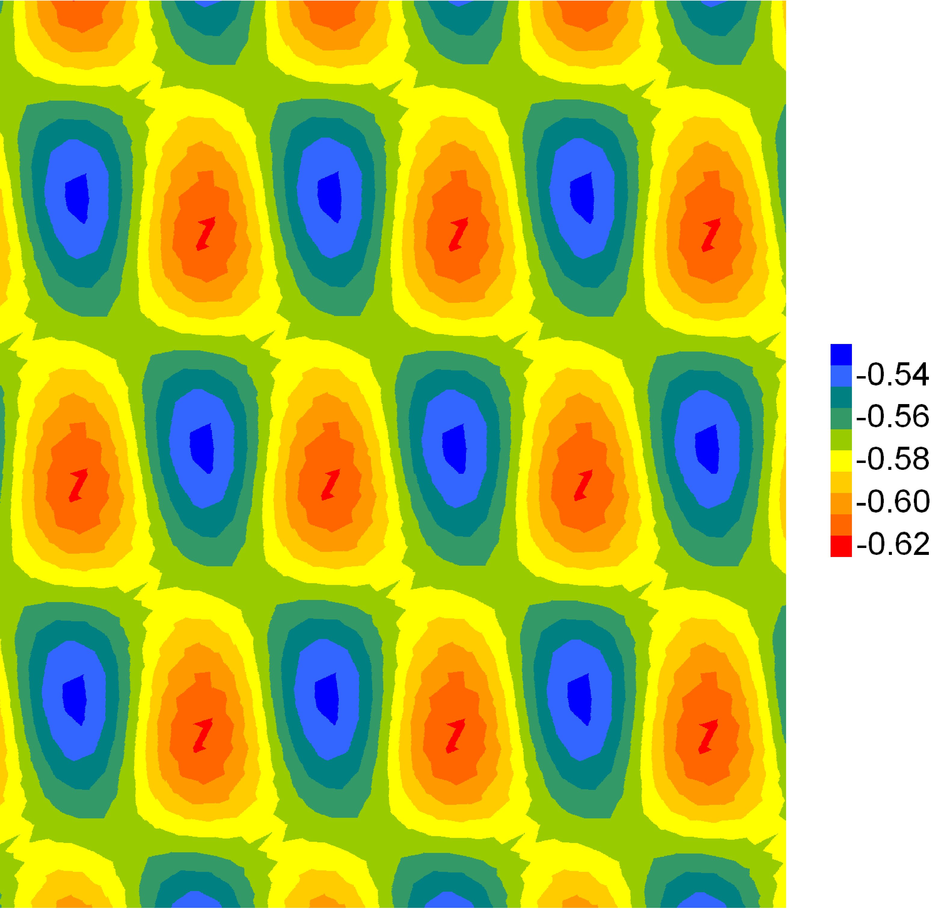 Figure 1
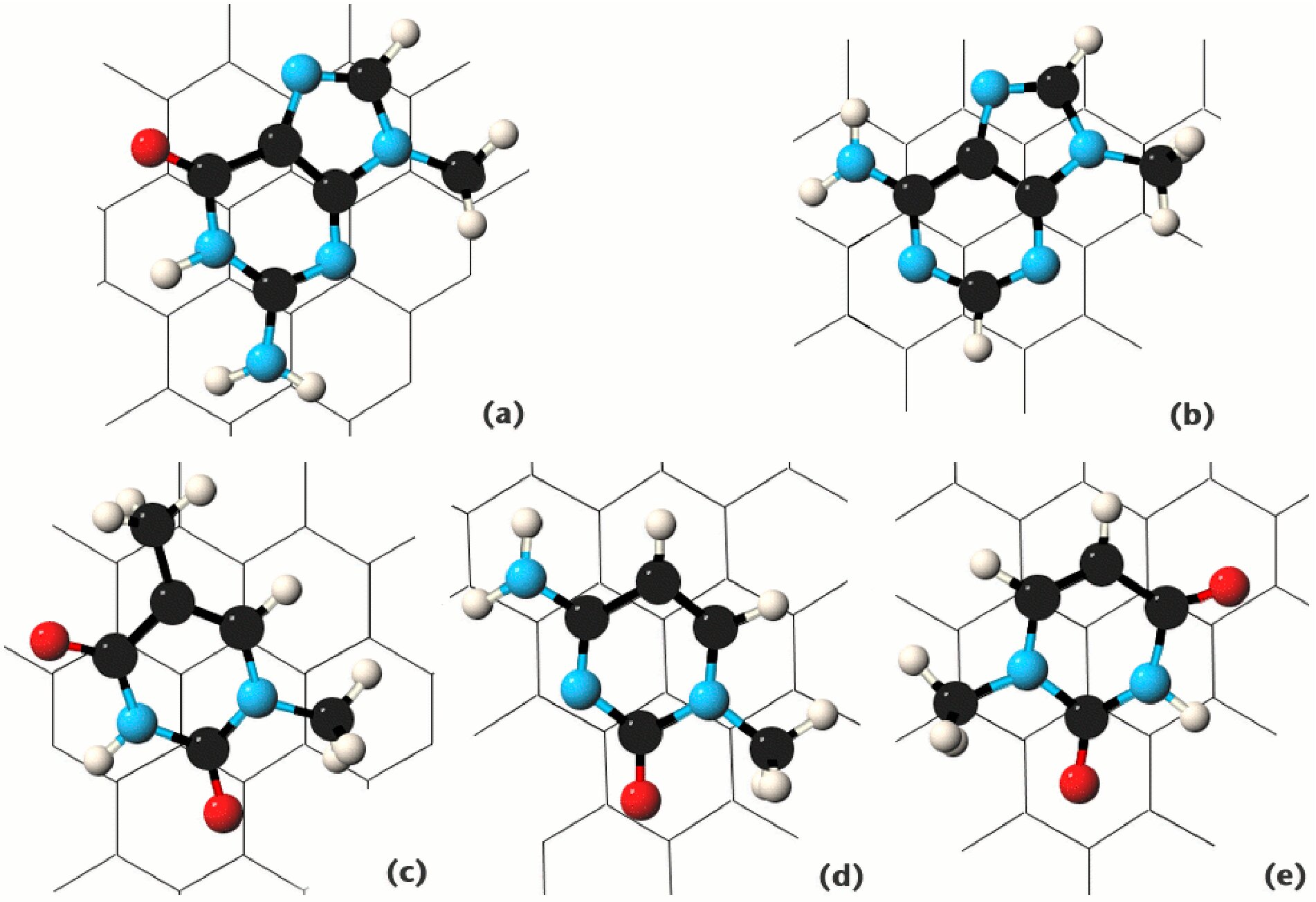 Figure 2
| base | (LDA) [eV] | (MP2) [eV] | [] |
|---|---|---|---|
| G | 0.61 | 1.07 | 131.2 |
| A | 0.49 | 0.94 | 123.7 |
| T | 0.49 | 0.83 | 111.4 |
| C | 0.49 | 0.80 | 108.5 |
| U | 0.44 | 0.74 | 97.6 |
Peer Reviews
No public reviews on file for this paper yet. If you reviewed it on a platform where reviews are public (OpenReview, ICLR, NeurIPS, ICML), you can paste yours below so the community can read it here.
Videos
No videos yet. Explain this paper in a talk, walkthrough, or lecture? Add one.
Physisorption of Nucleobases on Graphene
S. Gowtham1
Ralph H. Scheicher1,2
Rajeev Ahuja2,3
Ravindra Pandey1
[email protected], [email protected]
Shashi P. Karna4
1Department of Physics and Multi-Scale Technologies Institute, Michigan Technological University, Houghton, Michigan 49931, USA
2Condensed Matter Theory Group, Department of Physics, Box 530, Uppsala University, S-751 21 Uppsala, Sweden
3Applied Materials Physics, Department of Materials and Engineering, Royal Institute of Technology (KTH), S-100 44 Stockholm, Sweden
4US Army Research Laboratory, Weapons and Materials Research Directorate, ATTN: AMSRD-ARL-WM; Aberdeen Proving Ground, Maryland 21005-5069, USA
Abstract
We report the results of our first-principles investigation on the interaction of the nucleobases adenine (A), cytosine (C), guanine (G), thymine (T), and uracil (U) with graphene, carried out within the density functional theory framework, with additional calculations utilizing Hartree–Fock plus second–order Møller–Plesset perturbation theory. The calculated binding energy of the nucleobases shows the following hierarchy: G T C A U, with the equilibrium configuration being very similar for all five of them. Our results clearly demonstrate that the nucleobases exhibit significantly different interaction strengths when physisorbed on graphene. The stabilizing factor in the interaction between the base molecule and graphene sheet is dominated by the molecular polarizability that allows a weakly attractive dispersion force to be induced between them. The present study represents a significant step towards a first-principles understanding of how the base sequence of DNA can affect its interaction with carbon nanotubes, as observed experimentally.
pacs:
68.43.-h, 81.07.De, 82.37.Rs
DNA-coated carbon nanotubes represent a hybrid system which unites the biological regime and the nanomaterials world. They possess features which make them attractive for a broad range of applications, e.g., as an efficient method to separate carbon nanotubes (CNTs) according to their electronic properties Nakashima:2003 ; Zheng:2003a ; Zheng:2003b , as highly specific nanosensors, or as an in vivo optical detector for ions. Potential applications of single-stranded DNA (ssDNA) covered CNTs range from electron sensing of various odors Johnson:2005 , to probing conformational changes in DNA triggered by shifts in the surrounding ionic concentration Strano:2006a , and detection of hybridization between complementary strands of DNA Star:2006 ; Strano:2006b . The interaction of DNA with CNT is not limited to the outer surface of the tube; it has also been experimentally demonstrated that ssDNA can be inserted into a CNT Okada:2006 , further enhancing the potential applications of this nano-bio system.
The details of the interaction of DNA with CNTs have not yet been fully understood, though it is generally assumed to be mediated by the -electron networks of the base parts of DNA and the graphene-like surface of CNTs. One would like to obtain a better understanding of the binding mechanism, and the relative strength of base-CNT binding as it is indicated experimentally from sequence-dependent interactions of DNA with CNTs Zheng:2003b ; Johnson:2005 . In this Letter, we present the results of our first-principles study of the interaction of nucleobases with a graphene sheet as a significant step towards a deeper understanding of the interaction of ssDNA with CNTs.
Previous theoretical studies focused on the adsorption of the nucleobase adenine on graphite Ortmann:2005 . In the present study, we have considered all five nucleobases of DNA and RNA, namely the two purine bases adenine (A) and guanine (G), and the three pyrimidine bases cytosine (C), thymine (T), and uracil (U). Our specific interest is to assess the subtle differences in the adsorption strength of these nucleobases on graphene, which in turn will allow us to draw conclusions for the interaction of DNA and RNA with CNTs as well.
Calculations were performed using the plane-wave pseudopotential approach within the local density approximation (LDA) LDA ; LDA-remark of density functional theory (DFT) DFT , as implemented in the Vienna Ab-initio Simulation Package (vasp) VASP . The cutoff energy was set to 850 eV. For -point sampling of the Brillouin zone we used the Monkhorst-Pack grid Monkhorst:1976 , which we found from benchmark calculations to yield identical results as a Monkhorst-Pack grid would.
A array of the graphene unit cell in the - plane and a separation of 15 Å between adjacent graphene sheets in the -direction was found to be a suitable choice to represent the supercell. The base molecules were terminated at the cut bond to the sugar ring with a methyl group in order to generate an electronic environment in the nucleobase more closely resembling the situation in DNA and RNA rather than that of just individual isolated bases by themselves. This has the additional benefit that a small magnitude of steric hindrance can be expected from the methyl group, quite similar to the case in which a nucleobase with attached sugar and phosphate group would interact with graphene.
For each of the five nucleobases, an “initial force relaxation” calculation step determined the preferred orientation and optimum height of the planar base molecule relative to the graphene sheet. A slice of the potential energy surface was then explored by translating the relaxed base molecules in a fixed orientation parallel to the graphene plane in steps of 0.246 Å along the lattice vectors of graphene, covering its entire unit cell by a mesh of 10 10 scan points. The separation between base molecule and graphene sheet was held fixed at the optimum height determined previously. The determination of the minimum total energy configuration was then followed by a 360∘ rotation of the base molecules in steps of 5∘ to probe the dependence of the energy on the orientation of the base molecules with respect to the underlying 2-D graphene sheet. The configuration yielding the minimum total energy was used in the final optimization step in which all atoms in the system were free to relax. We would like to emphasize here that for all five nucleobases, the eventually determined equilibrium configuration was characterized by a separation between base and graphene sheet that was equal to the optimum height chosen in the previous lateral potential energy surface scan.
An additional set of calculations was performed using the ab initio Hartree–Fock approach coupled with second–order Møller–Plesset perturbation theory (MP2) as implemented in the gaussian 03 suite of programs GAUSSIAN . Due to the use of localized basis sets (rather than plane-wave), the system here consisted of the five nucleobases on top of a patch of nanographene Enoki:2000:2002 , i.e., a finite sheet containing 28 carbon atoms. The LDA optimized configuration and the 6-311++G(d,p) basis sets for C, H, N and O atoms were used for the MP2 calculations.
The first optimization step involving the “initial force relaxation” led to a configuration of all five nucleobases in which their planes are likewise oriented almost exactly parallel to the graphene sheet with a separation of about 3.5 Å, characteristic for – stacked systems pi-stacking . The interaction of the attached methyl group with the graphene sheet results in a very small tilt of the molecule, with angles less than 5∘.
The base is translated 2.461 Å along both graphene lattice unit vectors respectively (maintaining a constant vertical distance of 3.5 Å from the sheet, as determined in the previous step), and rotated 360∘ in the equilibrium configuration with respect to the configuration obtained after the “initial force relaxation” step in the optimization procedure. From the optimization steps involving the translational scan of the energy surface, it is apparent that the energy barriers to lateral movement of a given base can range from 0.04 to 0.10 eV (Fig. 1), thus considerably affecting the mobility of the adsorbed nucleobases on the graphene sheet at room temperature, and constricting their movement to certain directions. The rotational scans carried out by us found energy barriers of up to 0.10 eV, resulting in severe hindrance of changes in the orientation of the adsorbed nucleobase.
In their equilibrium configuration, three of the five bases tend to position themselves on graphene in a configuration reminiscent of the Bernal s AB stacking of two adjacent graphene layers in graphite (Fig. 2). Virtually no changes in the interatomic structure of the nucleobases were found in their equilibrium configurations with respect to the corresponding gas-phase geometries, as it could be expected for a weakly interacting system such as the one studied here. A notable exception is the RC-O in guanine which shows a 10% contraction upon physisorption of the molecule on graphene.
The stacking arrangement shown in Fig. 2 can be understood from the tendency of the –orbitals of the nucleobases and graphene to minimize their overlap, in order to lower the repulsive interaction. The geometry deviates from the perfect AB base-stacking as, unlike graphene, the six- and five-membered rings of the bases possess a heterogeneous electronic structure due to the presence of both nitrogen and carbon in the ring systems. In addition, there exist different side groups containing CH3, NH2, or O, all of which contribute to the deviation from the perfect AB base-stacking as well. Adenine, thymine and uracil display the least deviation from AB stacking (Fig. 2) out of the five nucleobases. For guanine and cytosine on the other hand, there is almost no resemblance to the AB stacking configuration recognizable (Fig. 2).
We calculated the binding energy for all five nucleobases. The binding energy of the system consisting of the nucleobase and the graphene sheet is taken as the energy of the equilibrium configuration with reference to the asymptotic limit obtained by varying the distance between the base and the graphene sheet in the -direction (Table 1). Within LDA, we found adenine, cytosine and thymine to all possess nearly identical binding energies of about 0.49 eV, while guanine with 0.61 eV is bound more strongly, and uracil with 0.44 eV somewhat more weakly.
It is somewhat surprising that guanine and adenine would possess such different physisorption energies, despite both containing a five- and a six-membered ring and featuring relatively similar molecular structures. A closer analysis of the various contributions to the total energy (Fig. 3) reveals that the Kohn-Sham kinetic energy displays a slightly more pronounced minimum for guanine than for adenine, and that the position of that minimum is shifted by about 0.25 Å towards the graphene sheet. The exchange-correlation energy drops off somewhat more rapidly in the case of adenine; however, the difference to the case for guanine is only very small.
Table 1 also includes the polarizabilities of the nucleobases calculated at the MP2 level of theory. The polarizability of the nucleobase Seela:2005 , which represents the deformability of the electronic charge distribution, is known to arise from the regions associated with the aromatic rings, lone pairs of nitrogen and oxygen atoms. Accordingly, the purine base guanine appears to have the largest value, whereas the pyrimidine base uracil has the smallest value among the five nucleobases. Our calculations confirm this behavior.
A remarkable correlation is found when the molecular polarizabilities of the base molecules are compared with the binding energies, in particular when the latter are also determined at the MP2 level of theory (Table 1). Clearly, the polarizability of a nucleobase is the key factor which governs the strength of interaction with the graphene sheet. This behavior is expected for a system that draws its stabilization from van der Waals (vdW) dispersion forces, since the vdW energy is proportional to the polarizabilities of the interacting entities. The observed correlation thus strongly suggests that vdW interaction is indeed the dominant source of attraction between graphene and the nucleobases.
The MP2 binding energies are systematically larger than those calculated within the LDA approximation (Table 1). This is due to the well established fact that MP2 provides a more accurate treatment of the vdW interaction than LDA. We note that the adsystem consisting of the base and the sheet is not bound at the Hartree-Fock level of theory, which underscores the importance of electron correlation in describing the weak vdW interactions in this system.
In the equilibrium configuration, a redistribution of the total charge density within a given base seems to appear. From an analysis of the Mulliken charges for the MP2 calculations, we also find a negligible charge transfer ( 0.02 e) between any of the five nucleobases and patch of nanographene in the equilibrium configuration. Electrostatic interactions in the adsystem are therefore very unlikely to contribute to the interaction energy.
In summary, we investigated the physisorption of the five DNA/RNA nucleobases on a planar sheet of graphene. Our first-principles results clearly demonstrate that the nucleobases exhibit significantly different interaction strengths when physisorbed on graphene. This finding represents an important step towards a better understanding of experimentally observed sequence-dependent interaction of DNA with CNTs Zheng:2003b ; Johnson:2005 . The calculated trend in the binding energies strongly suggests that the polarizability of the base molecules determines the interaction strength of the nucleobases with graphene. As graphene can be regarded as a model system for CNTs with very small surface curvature, our conclusions should therefore also hold for the physisorption of nucleobases on large-diameter CNTs. Further studies involving the investigation of nucleobases interacting with small-diameter CNTs are currently underway.
The authors acknowledge helpful discussions with Prof. Roberto Orlando of the University of Turin, Italy, and with Dr. Takeru Okada and Prof. Rikizo Hatakeyama of Tohoku University, Japan. S.G., R.H.S., and R.P. would like to thank DARPA for funding. R.H.S. and R.A. are grateful to the Swedish National Infrastructure (SNIC) for computing time. R.H.S. acknowledges support from EXC!TiNG (EU Research and Training Network) under contract HPRN-CT-2002-00317. The research reported in this document was performed in connection with contract DAAD17-03-C-0115 with the U.S. Army Research Laboratory.
The reference list from the paper itself. Each links out to its DOI / PubMed record.
- 1(1) N. Nakashima et al. , Chem. Lett. 32 , 456 (2003).
- 2(2) M. Zheng et al. , Nature Mater. 2 , 338 (2003).
- 3(3) M. Zheng et al. , Science 302 , 1545 (2003).
- 4(4) C. Staii et al. , Nano Lett. 5 , 1774 (2005).
- 5(5) D. A. Heller et al. , Science 311 , 508 (2006).
- 6(6) A. Star et al. , Proc. Natl. Acad. Sci. U.S.A. 103 , 921 (2006).
- 7(7) E. S. Jeng et al. , Nano Lett. 6 , 371 (2006).
- 8(8) T. Okada et al. , Chem. Phys. Lett. 417 , 288 (2006).